Rolando Pinho
MD
Endoscopy Unit Clínica Radelfe - Paços de Ferreira, Portugal
PATIENT INFORMATION / INDICATION
A 65 years old male was referred for colonoscopy after a positive fecal immunochemical test.
METHODS & RESULTS
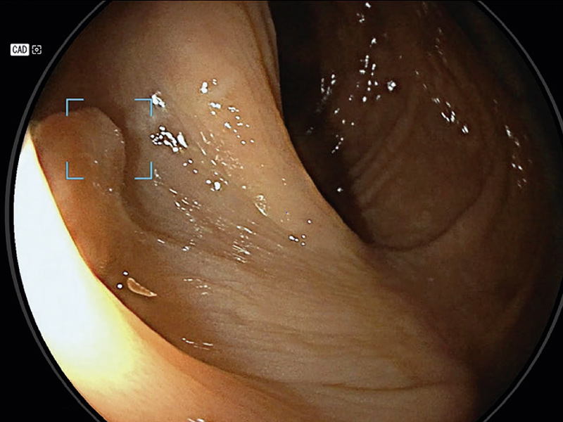
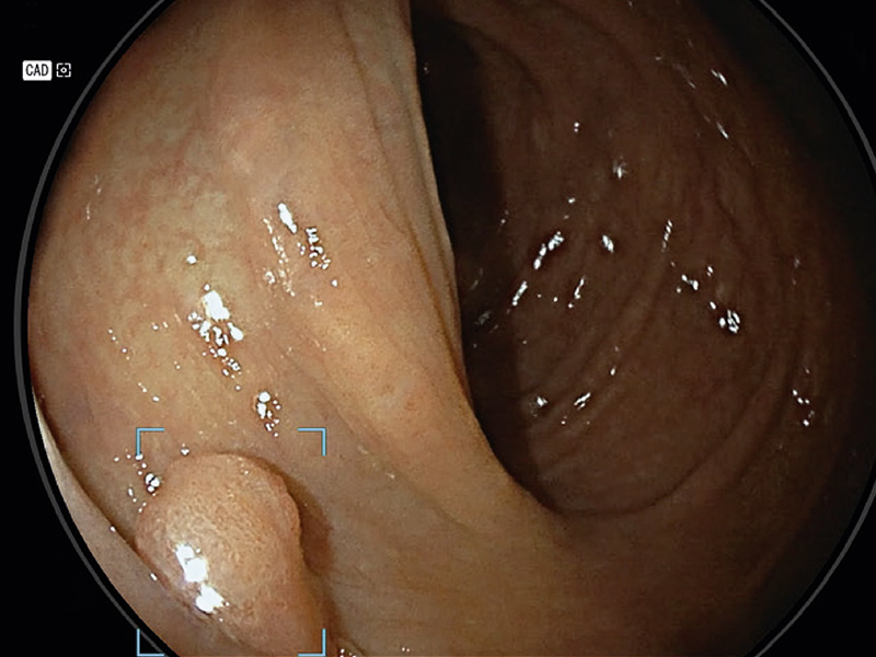
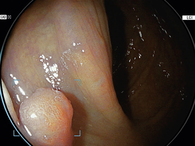
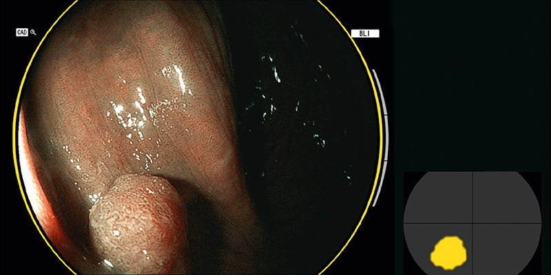
A small polyp hidden between two folds was detected by CAD-EYE (figure 1) in the sigmoid colon. After detection, the polyp was properly evaluated under White Light Imaging (WLI) (Figure 2) and Linked Color Imaging (LCI) (Figure 3). After changing to the Blue Light Imaging (BLI) mode, the CAD-EYE system classified the polyp as a neoplastic polyp (Figure 4).
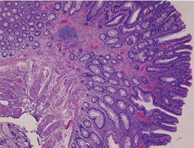
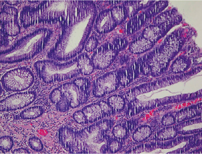
CONCLUSION
After polypectomy, histological analysis demonstrated the presence of a tubulovillous adenoma with low-grade dysplasia (Figure 5, 6).