Dr Dimitrios Karagiannis
MD PhD
Endoscopy Unit Athens Medical Center, Greece
PATIENT INFORMATION / INDICATION
A 61 years old woman with osteoporosis underwent a routine follow-up colonoscopy after a previous polypectomy five years ago (2015).
METHODS & RESULTS
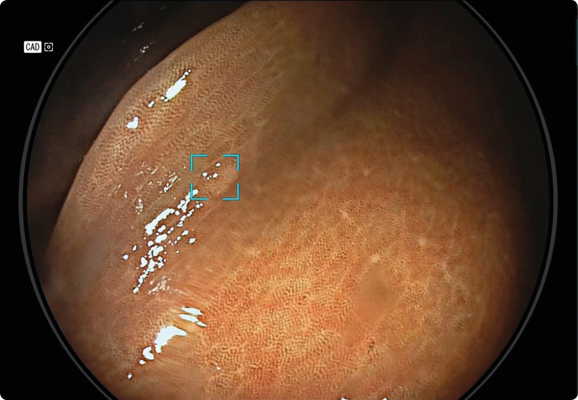
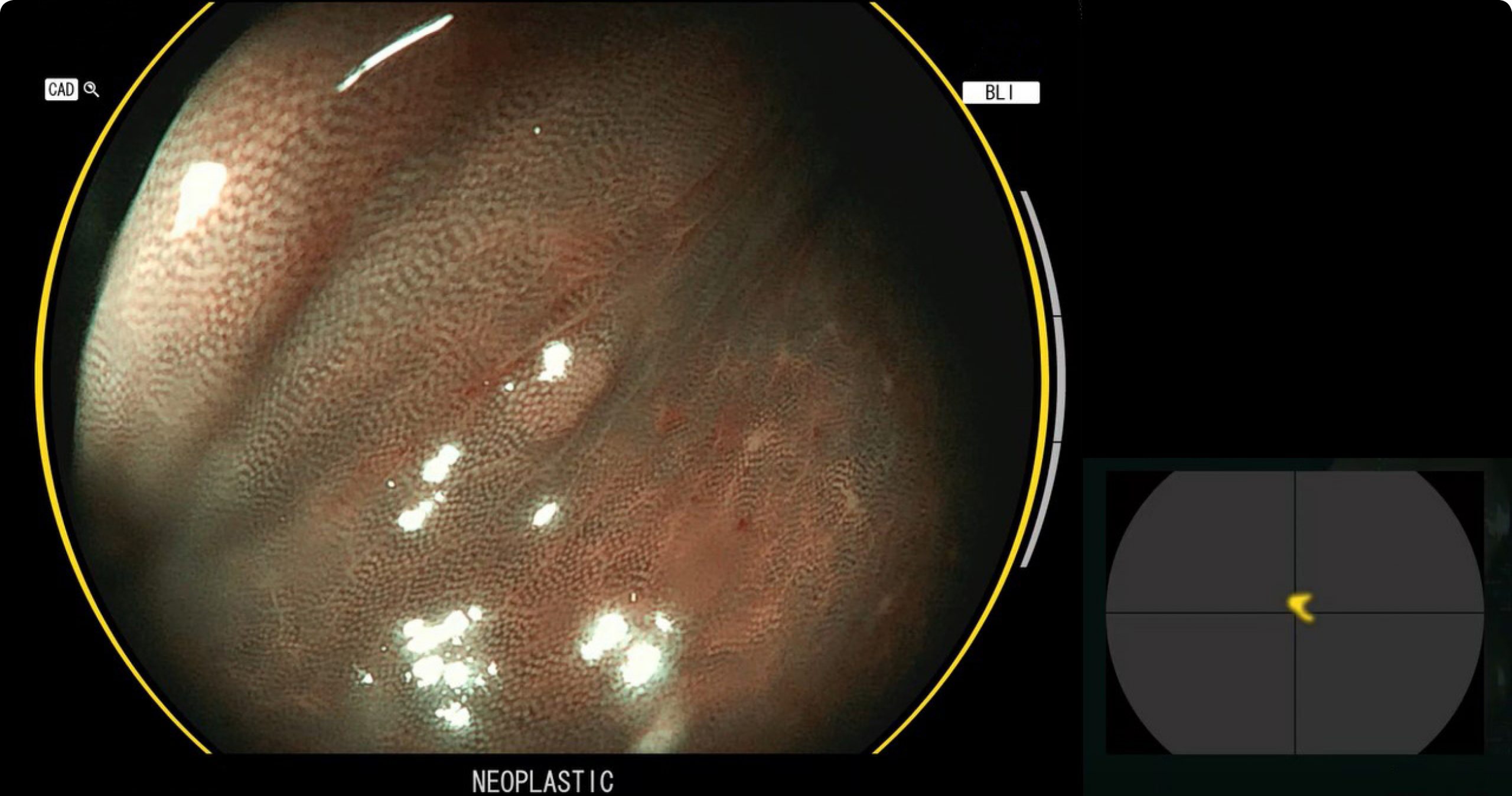
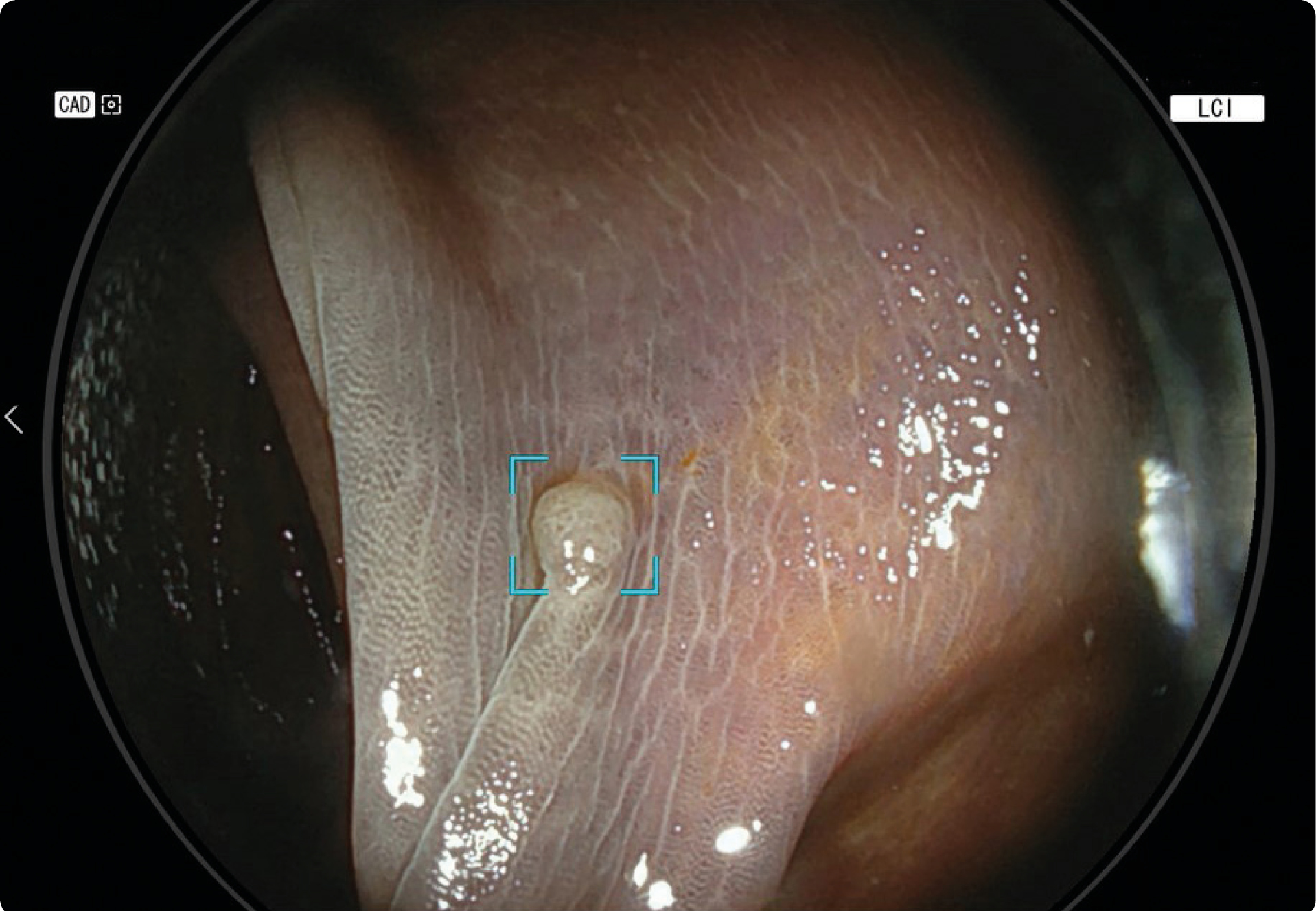
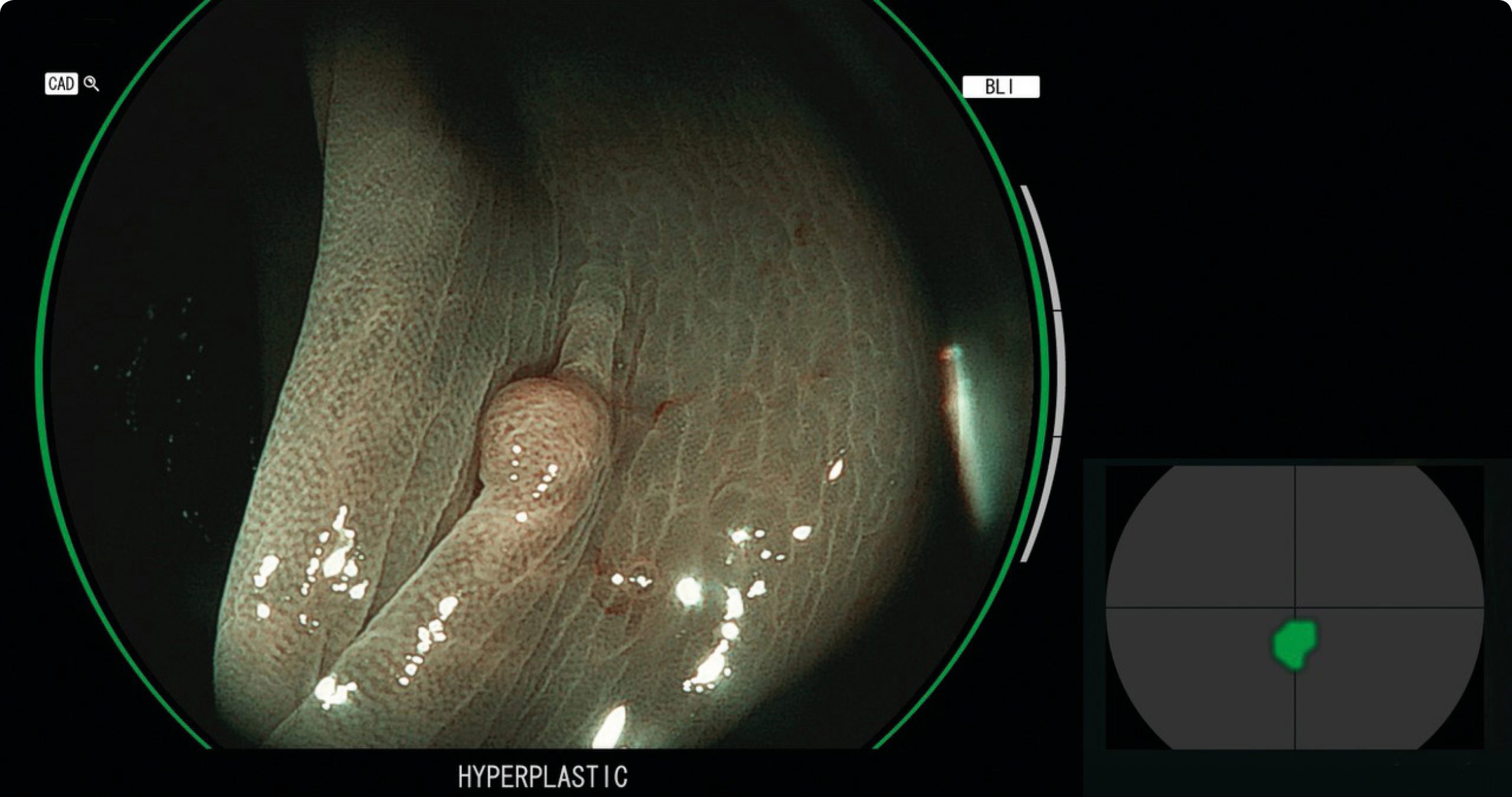
In this case we had a patient with melanosis coli, a brown discoloration of colonic mucosa, in which CAD EYE Detection and Characterisation performed extremely well. In this melanosis environment, a small polyp was detected by CAD EYE with White Light in the right flexure (Fig.1a) which was predicted with Blue Light Imaging (BLI) as adenoma (Fig.1b). Within the folds of the transverse colon, a second small polyp was detected with Linked Color Imaging (LCI) (Fig. 2a) which CAD EYE diagnosed as hyperplastic (Fig.2b). Both polyps were removed and sent for pathological analysis.
CONCLUSION
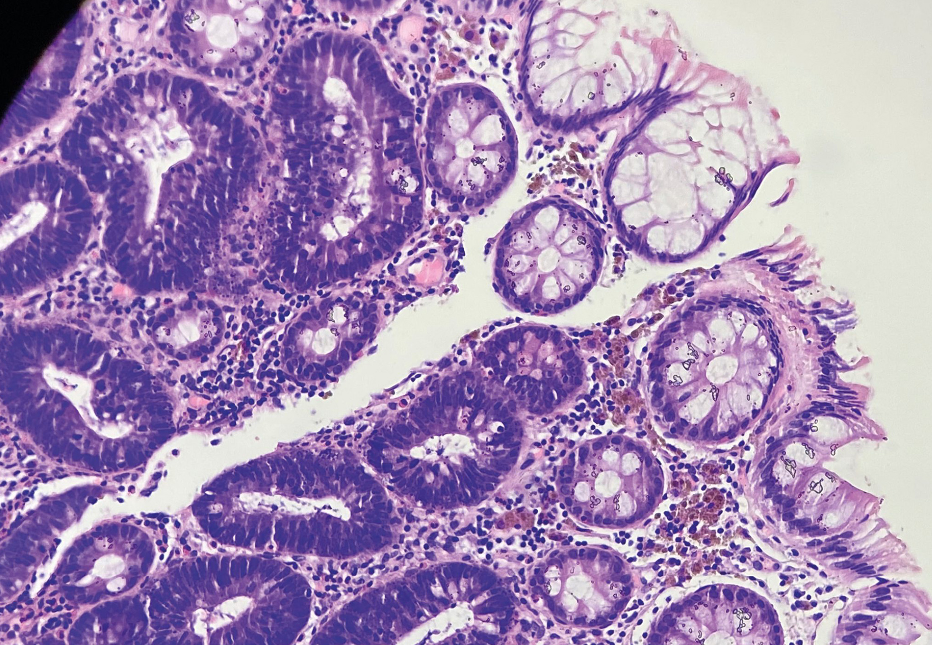
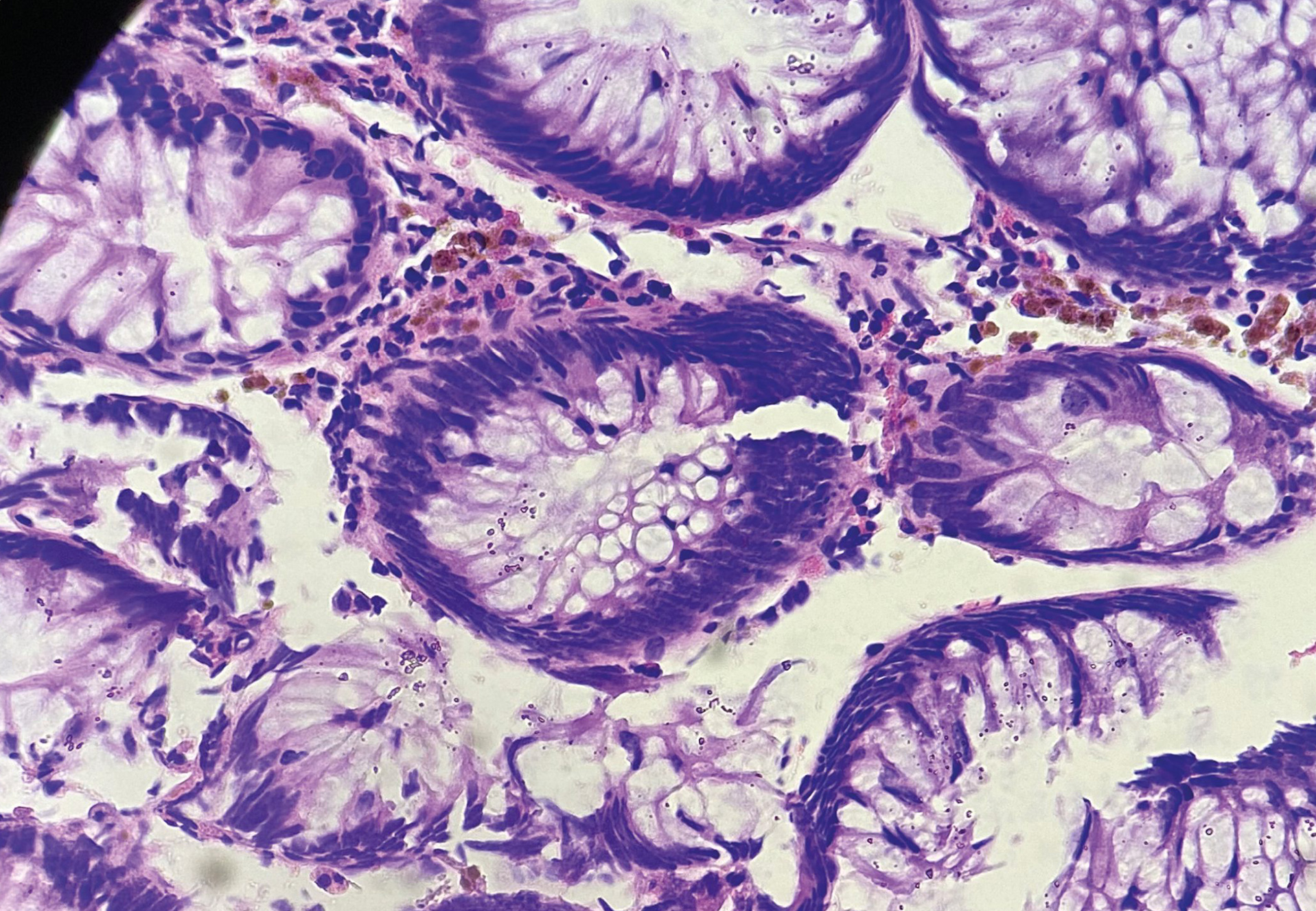
The pathological results by Dr. C. Spiliadi confirmed the prediction by CAD EYE for both polyps (Fig.1c and Fig.2c):
Fig.1c Adenoma with low grade dysplasia on melanosis coli due to laxatives.
Fig.2c Hyperplastic polyp, no dysplasia on melanosis coli due to laxatives.