Emanuele Rondonotti
MD PhD
Ospedale Valduce Gastroenterology Unit Como, Italy
PATIENT INFORMATION / INDICATION
A 70 years old male underwent faecal immunochemical blood test, as part of the Regional Colorectal Cancer screening programme, with positive result. He was therefore referred for colonoscopy.
METHODS & RESULTS
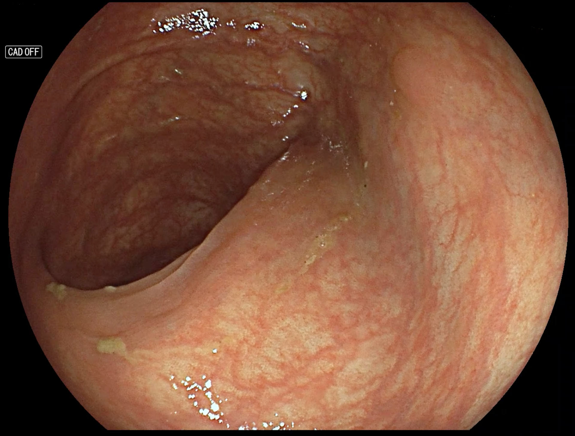
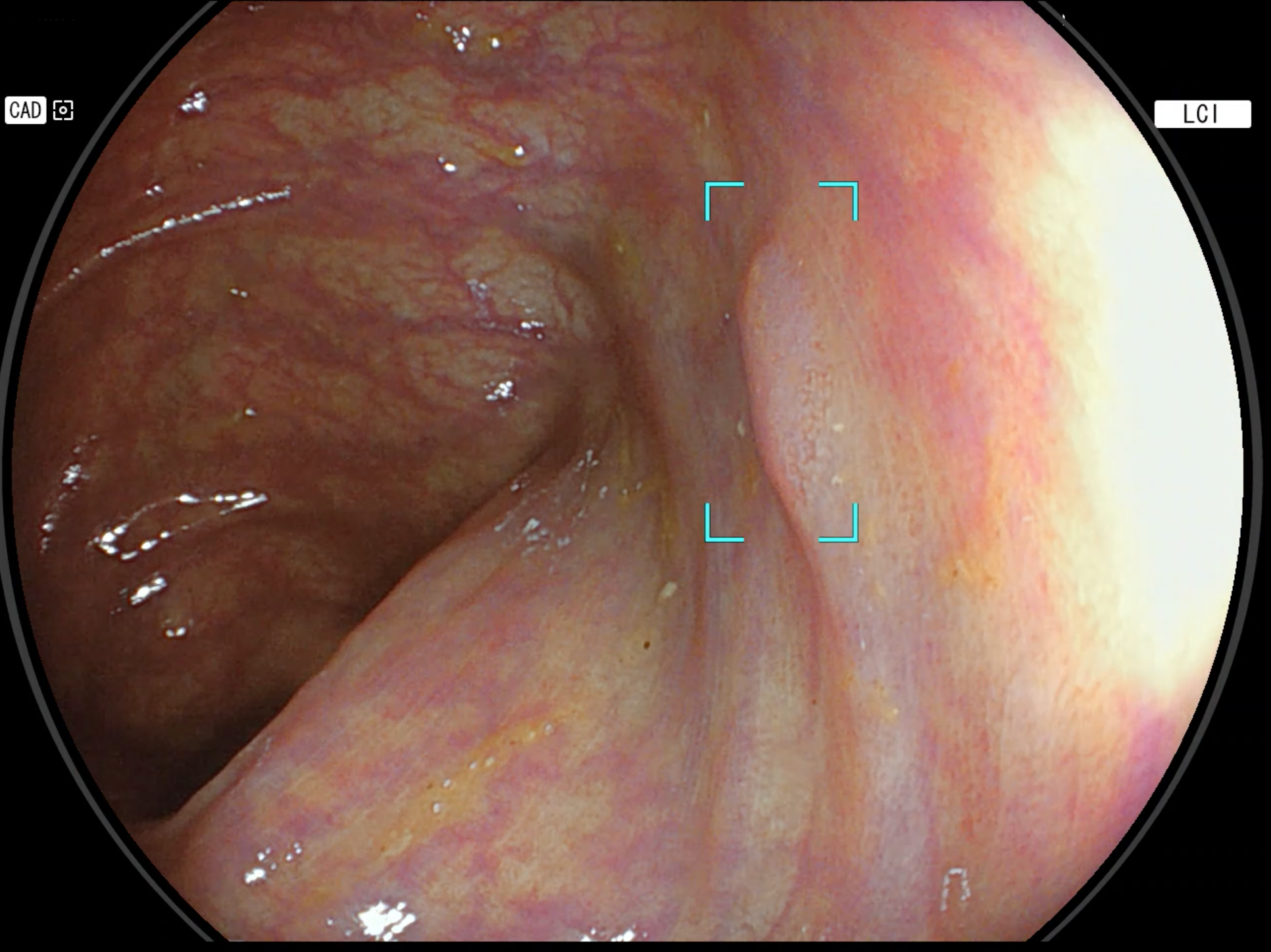
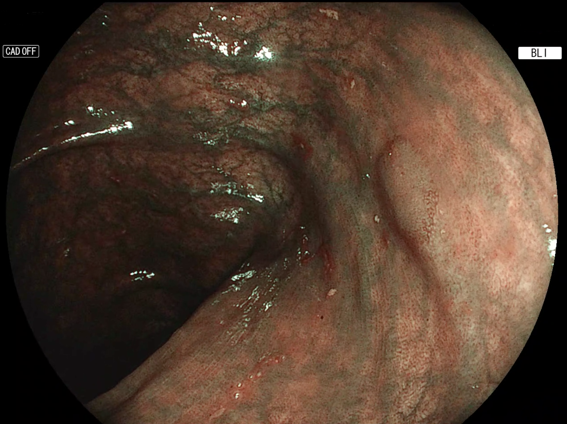
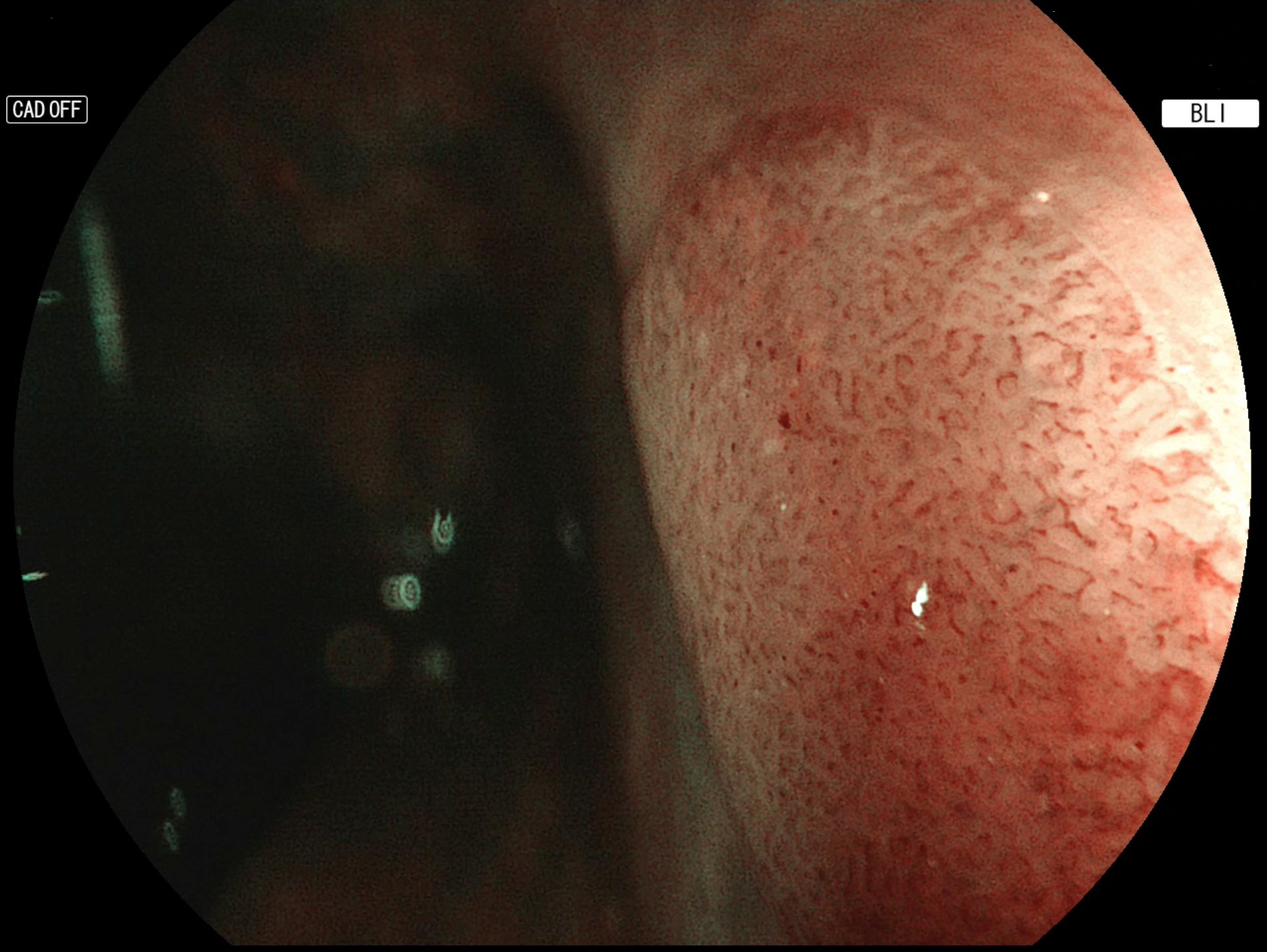
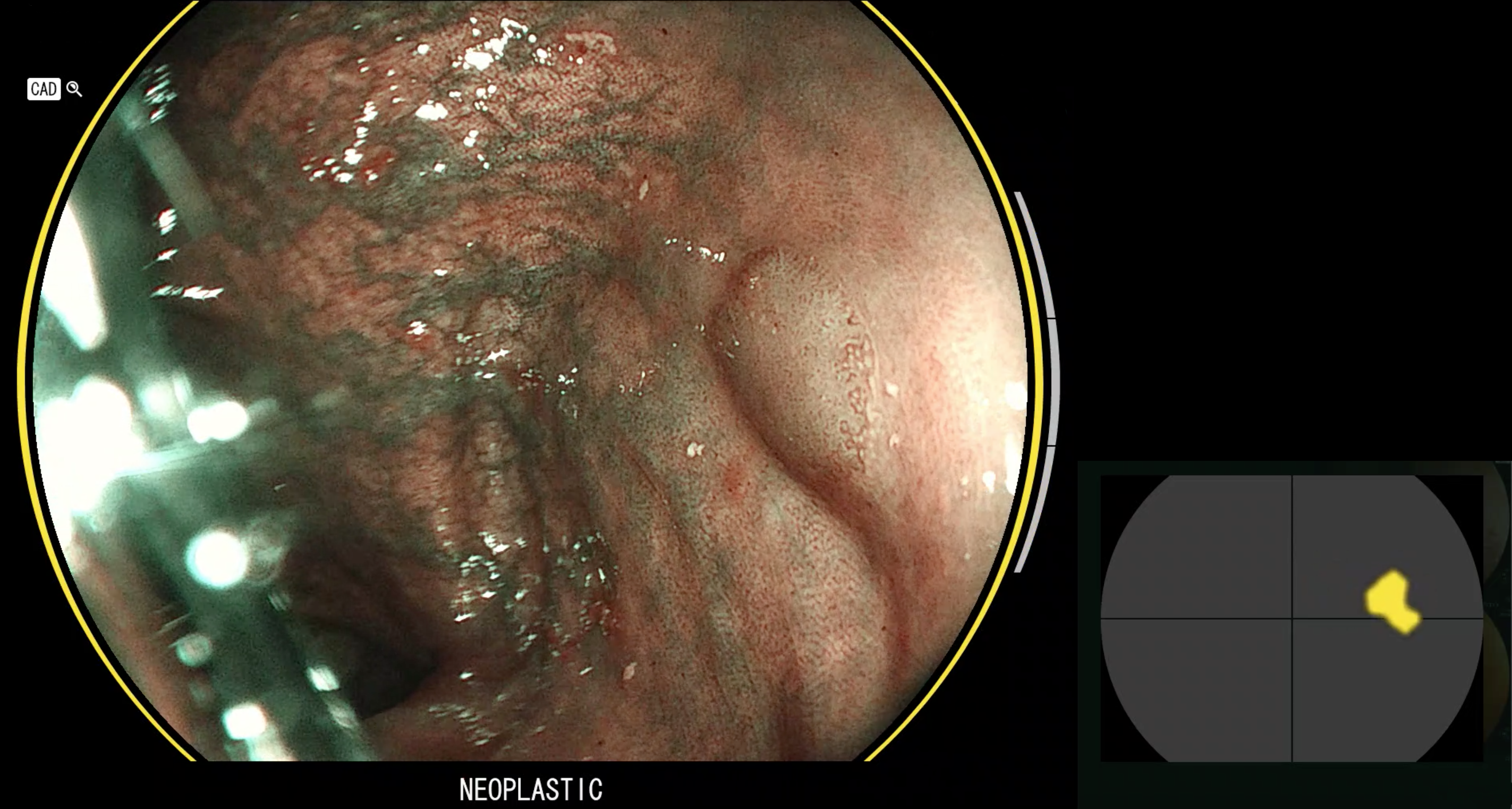
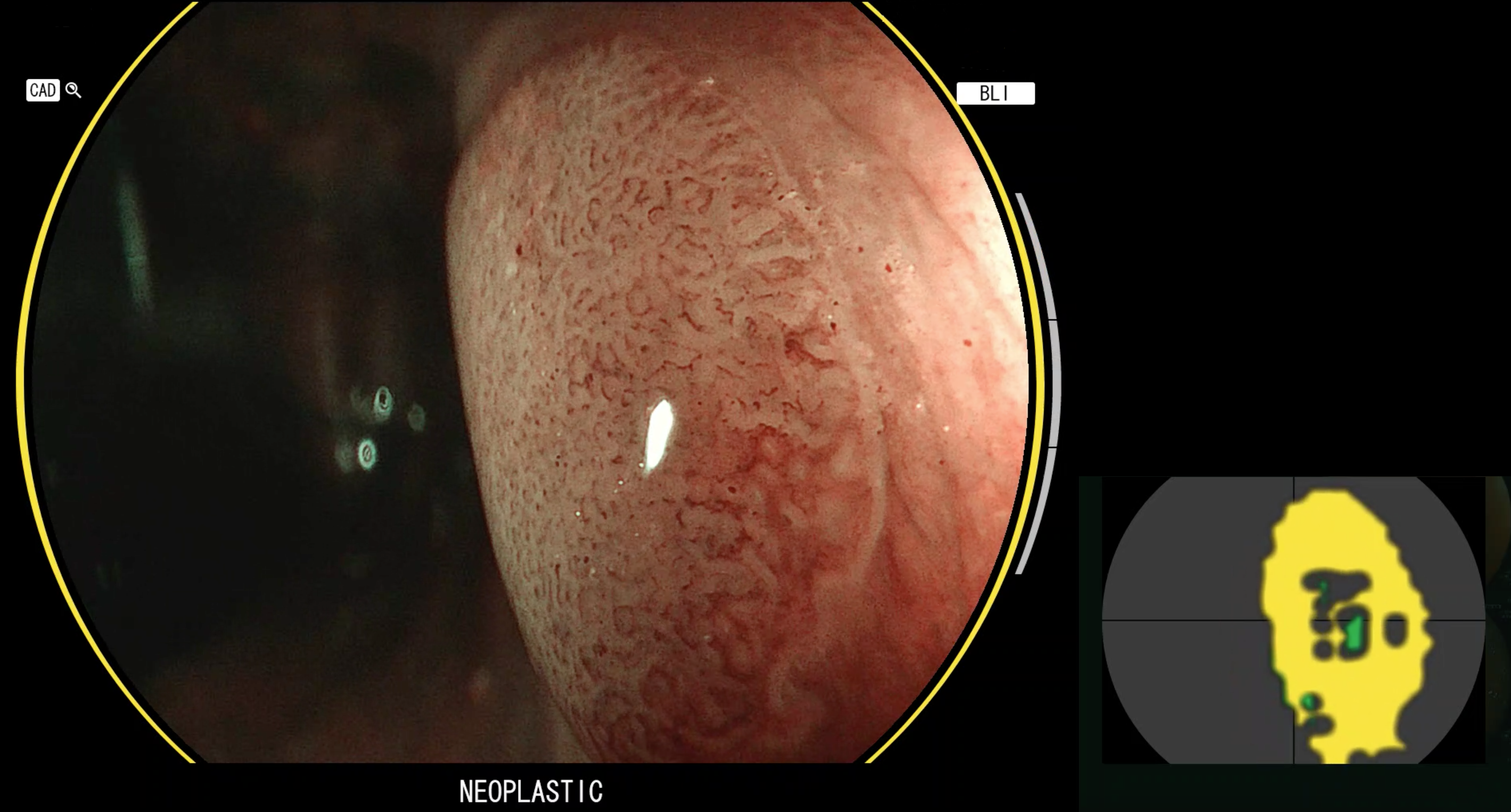
During colonoscope withdrawal, two diminutive sessile polyps were detected in the ascending colon, while a 5 mm flat lesion was identified in the sigmoid. This was barely visible with White Light (Figure 1), whereas it was easily identified by combining Linked Color Imaging (LCI) and CAD EYE (Figure 2). In order to real-time characterise this lesion, the Blue Light Imaging (BLI) mode (Figure 3) was then activated. Unfortunately, the polyp was difficult to categorise, according to BASIC (BLI Adenomatous Serrated International Classification) criteria: it showed superficial features typical of both hyperplastic and adenomatous polyps. Neither the use of magnification nor a closer and more detailed observation (Figure 4) allowed to confidently predict its histology; the polyp was then classified by the endoscopist as an adenoma, but with low confidence.
When the CAD EYE for polyp characterisation was switched on, the polyp was consistently characterised as adenoma (Figure 5); this was also confirmed after image magnification (Figure 6). Since the endoscopist’s initial assessment was consistent with the output provided by the system, he stated that he could finally classify the polyp with high confidence as adenoma.
CONCLUSION
Histological analysis revealed a tubular adenoma with low-grade dysplasia. In such a case, the CAD EYE system may assist the endoscopist and facilitate the histology prediction process.