Emmanuel Coron
Prof. Dr.
Digestive Diseases Institute, University Hospital of Nantes, France
A slightly-elevated flat lesion (Paris 0-IIa) was found in the middle of a Barrett’s oesophagus. LCI allowed easier detection of the lesion which consisted in several nodules with an irregular vascular pattern. The final histology was high-grade dysplasia with p53 overexpression (Figs. 1 + 2).
A second slightly-elevated flat lesion (Paris 0-IIa) was found in the same patient. LCI allowed easier detection of the lesion, which was more whitish that the surrounding mucosa and with clearer delineation of margins. The final histology was high-grade dysplasia with no overexpression of p53 (Figs. 3 + 4).
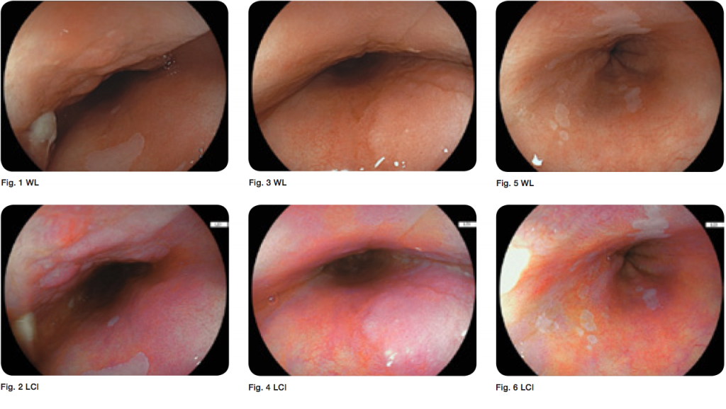
The lower part of this Barrett’s oesophagus showed several patches of normal squamous mucosa, which were easily detected by both WLE and LCI. LCI also showed reddish areas corresponding to inflamed tissue, which was subsequently confirmed by biopsy (incomplete intestinal metaplasia, with moderate inflammation) (Figs. 5 + 6).