Prof. Dr. Evelien Dekker
MD
Gastroenterology & Hepatology, Academic Medical Center, University of Amsterdam, Netherlands
Patient information / Indication
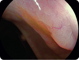
71-year old woman with serrated polyposis syndrome.
Methods & Results
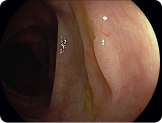
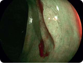
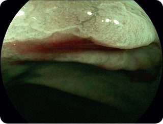
During this colonoscopy 12 polyps were removed. Except for 1 tubular adenoma, all of these were serrated polyps. The pictured polyp is a sessile serrated lesion (Figure 1). Using BLI (Figure 2) and BLI zoom (Figure 3), these polyps can be differentiated using the WASP-criteria (Joep et al., GUT 2016): dark spots inside crypts and irregular shape. In this case, however, the borders are remarkably well defined. BLI also shows a clear ‘red cap sign’ (Saito et al., World J Gastrointest Endosc. 2015), owing to the mucus that frequently cover or surround sessile serrated lesions
Conclusion
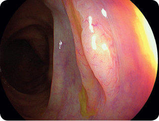
Using LCI zoom (Figure 4 + 5), the characteristic cloudy surface is clearly visualised.