Prof. Pradeep Bhandari
MD
Solent Centre for Digestive Diseases, Queen Alexandra Hospital Portsmouth, UK
Patient information / Indication
An 80-year old man was referred for endoscopic mucosal resection of a 10 cm rectal adenoma. His main symptom was debilitating mucous discharge and diarrhoea. He had multiple cardiac co-morbidities that prevented curative surgical resection. The endoscopic assessment was performed to exclude foci of cancer.
Methods & Results
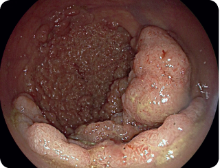
By using the Fujifilm 700 series gastroscope (EG-760R) the extensive adenoma was visualised encompassing 100 % of the luminal circumference.
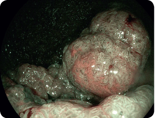
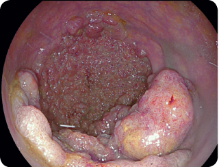
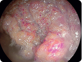
Figure 1 is the white light image. BLI did not reveal any evidence of invasive vascular pattern (Figure 2). LCI highlighted the villous nature of the polyp (Figure 3, 4).
Conclusion
Giant polyps have a high risk of cancer and are usually not suitable for endoscopic resection. BLI assessment allowed us to confidently exclude malignancy in this case and consider endoscopic resection.